
Figure 6.
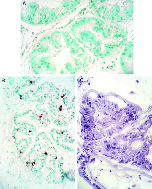
In situ detection of epithelial cells undergoing apoptosis by the ApopTag kit (Oncor). A: A representative section of the DLP from an untreated intact rat (mean AI value for the group, 0.9 ± 0.3%; n = 3 animals per group). No apoptotic cells are present in this section of DLP. In other sections and in glands from intact untreated rats, occasional staining of one or more glandular cells was found. Methyl green counterstain; magnification, ×220. B: DLP section from a rat castrated 7 days before its death. Numerous epithelial cells contain positively stained brown nuclei in these atrophic glands (mean AI value for the group, 11.0 ± 3.0%; n = 3 animals per group). There is considerable agreement here between the apoptotic activity and degree of TRPM-2/clusterin expression in this tissue. Methyl green counterstain; magnification, ×150. C: A DLP dysplastic lesion from a rat treated with T+E2 for 16 weeks. This section is from the same ductal lesion illustrated in Figures 4, A and B, and 5C ▶ ▶ . Note the piling up of dysplastic epithelial cells, some of which have formed pseudoacini, and the variations in size and shape of nuclei. No positively stained cells were detected in these dysplastic lesions. Compare the total absence of apoptosis in this section with the intense expression of TRPM-2/clusterin message and protein, illustrated in Figures 4, A and B, and 5C ▶ ▶ , respectively. Harris hematoxylin counterstain; magnification, ×400.