
Figure 1.
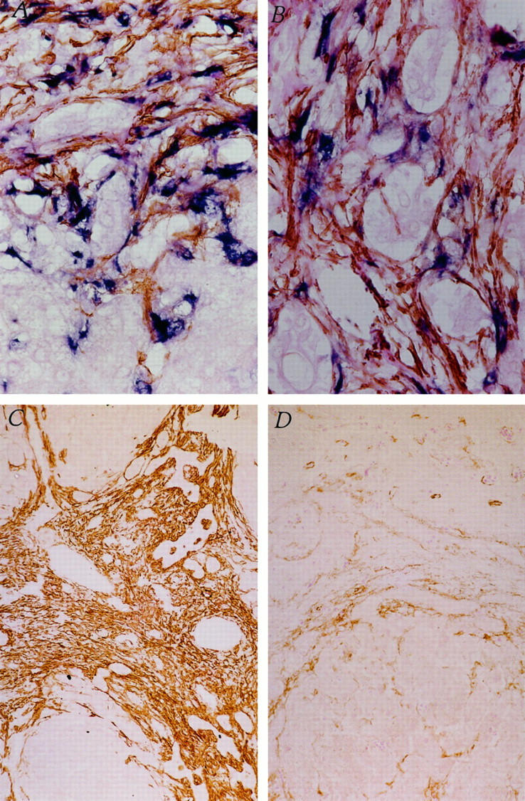
Identification of activated HSCs as cellular source of increased collagen production in biliary atresia. A: Colocalization of SMA (brown) and procollagen α1 (I) mRNA (blue) within stellate-shaped activated HSCs in a liver biopsy from an infant with biliary atresia, using immunohistochemistry and in situ hybridization, respectively. Original magnification, ×400. B: Bile duct hyperplasia within fibrotic bands in a liver biopsy from an infant with biliary atresia. Intense staining for SMA (brown) and procollagen α1 (I) mRNA (blue), colocalized in activated HSCs. Original magnification, ×400. C: Immunohistochemistry for SMA (brown) within activated HSCs surrounding hyperplastic bile ducts in a pre-Kasai HPE. D: A post-Kasai HPE liver biopsy from the same infant shown in C with biliary atresia. Original magnification, ×100.