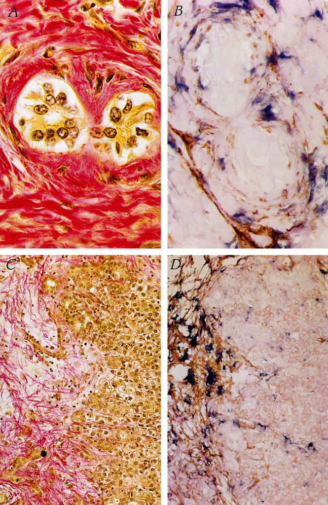
Figure 2.

Colocalization of activated HSCs to collagen protein deposition in biliary atresia liver. A: Collagen protein deposition surrounding two bile ducts in liver biopsy from an infant with biliary atresia (pink). B: Activated HSCs surrounding bile ducts, showing colocalization of SMA (brown) and procollagen α1 (I) mRNA (blue). Original magnification, ×1000. C and D: Fibrotic region in liver biopsy from an infant with biliary atresia showing deposition of collagen protein fibrils (C, pink) and activated HSCs (D), as evidenced by colocalization of SMA (brown) and procollagen α1 (I) mRNA (blue). Original magnification, ×200.
