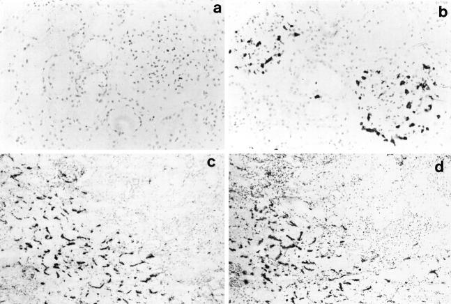
Figure 2.
Immunohistological changes observed after reperfusion in cadaver renal allografts. a: Indirect immunoperoxidase staining of a prereperfusion biopsy with an antineutrophil elastase mAb demonstrating the absence of neutrophils. b: The subsequent postreperfusion biopsy stained with the same antibody showing neutrophils infiltrating into the glomeruli (a and b, magnification, ×200). c: Postreperfusion biopsy stained by the indirect immunoperoxidase method using an anti-P-selectin antibody, showing a positive signal on the intertubular capillaries. d: A consecutive section from the same postreperfusion biopsy stained with the anti-CD41 platelet-specific antibody showing a pattern of staining similar to that detected for P-selectin (c and d, magnification, ×100).

