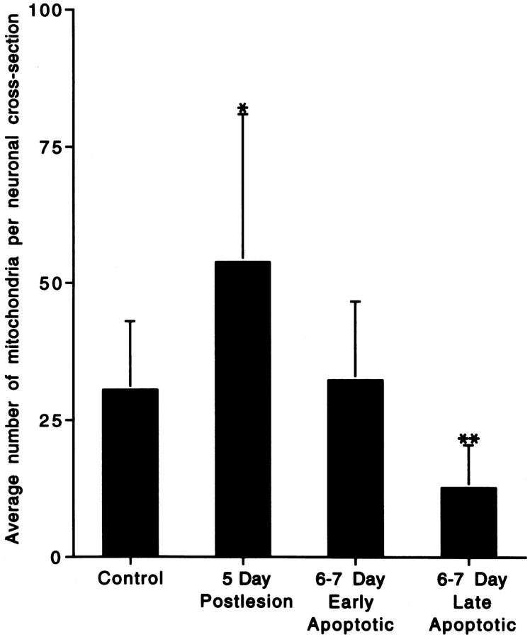
Figure 4.
Histogram of the semiquantitative analysis of mitochondrial accumulation within neuronal perikarya in the ventromedial dLGN after occipital cortex ablation. Values are means ± SD. At 5 days postlesion, the number of recognizable mitochondria per neuronal cross-section was 176% of control, whereas by 6 to 7 days postlesion, the number of recognizable mitochondria was 41% of control in neurons at late stages of apoptosis. *Significant difference (P = 0.07) as compared with control; **significant difference (P = 0.02 and P = 0.007) as compared with control and 5 days postlesion, respectively.

