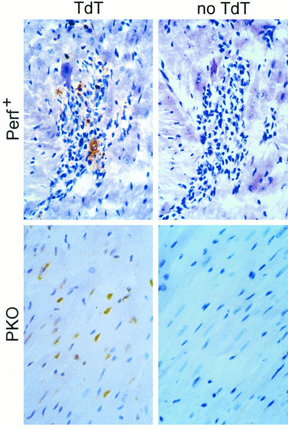
Figure 7.

Apoptosis in CVB3-infected myocardium. The hearts harvested at 10 days p.i. and evaluated for myocarditis (shown in Figure 3 ▶ ) were tested for apoptosis using the terminal deoxynucleotidyl transferase (TdT)-mediated nick-end labeling assay. Reactions were carried out in the presence of the enzyme TdT (left; TdT) or, as a control, in its absence (right; no TdT). Signal in perforin+ mice was seen only within the inflammatory foci (top left), whereas in PKO mice signal was absent from the small foci, but was detected in myocyte nuclei (bottom left).
