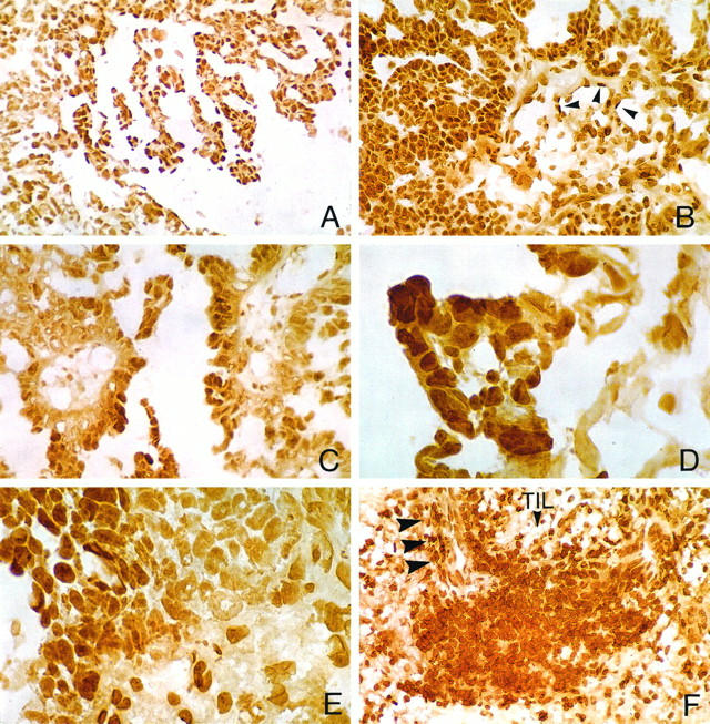
Figure 3.

Panel featuring differential cellular localization of RARα in serous adenocarcinomas. A to C: Widespread RARα immunohistochemical staining of neoplastic cells in well to moderately differentiated areas of grade II serous tumors with papillary morphology. B depicts, in addition, RARα staining of endothelial cells in vessels of the fibrovascular core (arrowheads). D and E illustrate, respectively, intratumoral staining heterogeneity associated with better differentiated, albeit abortive, RARα-positive papillary foci (D) or clusters of RARα-positive poorly differentiated tumor cells (E). F: Prominent nodular aggregate of TILs with robust RARα staining. Arrowheads point to a frequently observed perivascular predilection of RARα-positive TILs. Avidin-biotin complex peroxidase method without hematoxylin counterstaining; original magnifications: A and B, ×400; C to F, ×1000.
