
Figure 1.
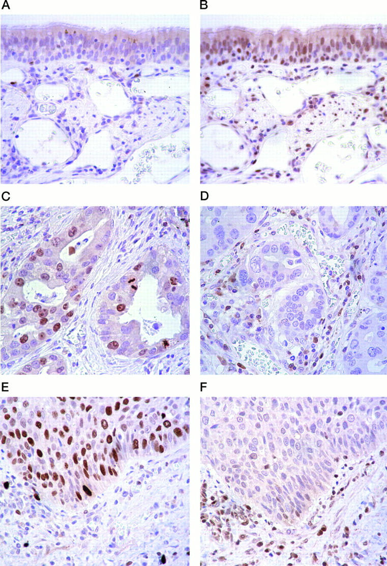
Comparison of Ki-67 antigen and p27 in nonneoplastic and cancerous lung tissues. Anti-Ki-67 antigen and anti-p27 immunostaining of sections of normal human lung tissue (A and B), adenocarcinoma (C and D), and squamous cell carcinoma (E and F) are compared. Ki-67 antigen expression was analyzed using monoclonal MIB-1 antibody. In normal lung tissue, immunostaining with MIB-1 showed few positive cells (A), whereas that with p27 showed positive nuclei in about 60 to 70% of cells (B). In adenocarcinoma tissue, immunostaining with MIB-1 revealed that 30% of cells had positive nuclei (C), whereas immunostaining of nuclei by anti-p27 showed few positive cells (D). In squamous cell carcinoma tissue, immunostaining with MIB-1 revealed positive nuclei in 60% of cells (E), whereas p27 immunostaining revealed few positive cells (F). Original magnification, ×400.