Figure 1.

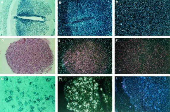
Expression of the SMN gene in fetal spinal cord. A to C: Spinal cord at 8 weeks of development. A: Untreated section, bright field. The cells from the neural epithelium migrate toward the posterior horn and, to a lesser degree, toward the anterior horn. B: Antisense probe, dark field, showing the diffuse expression in the neural epithelium following the pattern of migration of the neural cells into the two horns. C: Sense probe, dark field. Hematoxylin and eosin; (×100). D to F: Dorsal root ganglia at 12 weeks of development. D: Antisense probe, bright field. E: Antisense probe, dark field. F: Sense probe, dark field. Hematoxylin and eosin (×200). G to I: Anterior horn of the spinal cord at 17 weeks of development showing a large expression in neuroblasts. G: Antisense probe, bright field. TB (×400). H: Antisense probe, dark field. TB (×200). I: Sense probe. Hematoxylin and eosin (×200).
