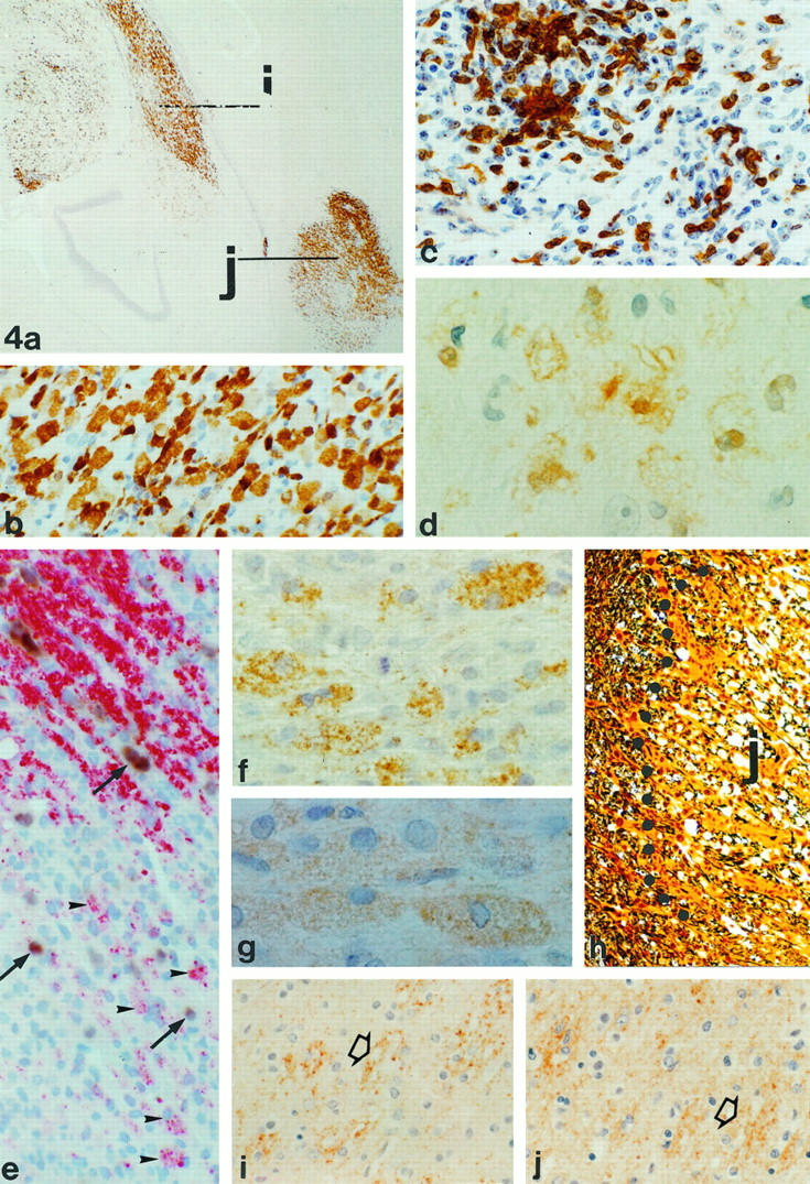
Figure 4.

Immunopathology of active lesions in monkey EI. a: Staining of regions I and J for macrophage marker 27E10 (magnification, ×34). Note that lesion I (see also Figures 2 and 3 ▶ ▶ ) shows 27E10 reactivity close to the lateral ventricle but also deeper in the parenchyma. b: Higher-magnification (×335) 27E10-positive macrophages in lesion J. c: Staining of CD3+ve cells in lesion I, indicating strong T-cell infiltration (magnification, ×335). d: MRP-14-positive macrophages in lesion I (magnification, ×990). e: Staining of lesion J with in situ hybridization for PLP mRNA combined with immunocytochemistry for PLP. The edge of this lesion shows the presence of macrophages containing PLP degradation products (arrowheads). Outside the lesion, in between the myelin (red), PLP mRNA-positive oligodendrocytes (brown; arrows) are found. In the lesion itself some weakly stained remaining oligodendrocytes (arrows) are present (magnification, ×388). f: PLP staining of lesion I showing the presence of macrophages with PLP degradation products (magnification, ×684). g: The presence of MOG degradation products in macrophages in lesion I (magnification, ×792). h: Bielschowsky silver impregnation for axons showing the edge of lesion J. Inside the lesion, likely because of edema, the density of axons is lower as in the surrounding tissue (magnification, ×161). Active demyelination in lesion I might occur through an antibody- and complement-mediated mechanism. i: Deposition of Igs on bundles of myelin (arrow). The punctate staining indicates that these myelin sheaths undergo degradation (magnification, ×308). j: The staining for complement C9 reveals that the same bundles that are opsonized with Igs also show deposition of C9 (magnification, ×308).
