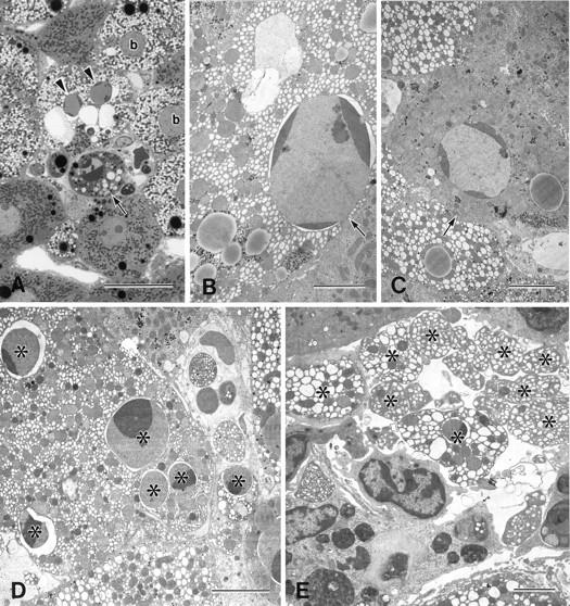
Figure 1.

Light and electron micrographs of rat liver 3 to 6 hours after the injection of CCl4. A: Ballooned hepatocytes (b) showed a pale, foamlike cytoplasm. Apoptotic hepatocytes with (B) or without (C) ballooned changes were observed in the lobule. Apoptotic foci in the midzonal area were composed of apoptotic hepatocytes with (arrowheads) and without (arrow) ballooned changes (A). Nuclear fragments (D*) and apoptotic bodies (E*), from the apoptotic ballooned cells were often observed. A, toluidine blue staining. Bars: A, 20 μm; B to E, 5 μm.
