
Figure 2.
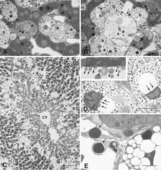
Light and electron micrographs of the liver 6 to 12 hours after the injection of CCl4. A: Necrotic cells (arrows) were found near the ballooned cells 6 hours after the injection of CCl4. B: A few ballooned hepatocytes (b, arrow) were undergoing necrosis. C: Ballooned cells encircled the centrilobular area 12 hours after the injection (cv, central vein). D: Nuclear blebbing (arrows) was observed in many of the ballooned cells, and the nuclei of the ballooned cells appeared crescent shaped. Marginated heterochromatin was apparent. D, inset: Higher magnification of the ballooned cell with nuclear blebbing, the blebs (arrowheads) protruded and detached from nucleus. E: The nuclei or nuclear fragments with highly condensed chromatin (arrows) found outside the cell were phagocytosed by the nearby cells. A and B, toluidine blue staining; C, hematoxylin and eosin staining. Bars: A and B, 20 μm; C, 80 μm; D, 10 μm; E, 2 μm.