
Figure 3.
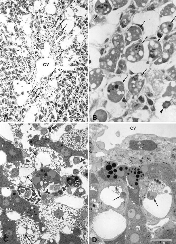
Light micrographs of the liver 24 to 72 hours after the injection of CCl4. A: Many centrilobular hepatocytes (arrows) were seen to undergo apoptosis (cv, central vein). B: Same image as in A, with a higher magnification. A ballooned hepatocyte (arrowhead) exhibited typical condensed chromatic masses in its nucleus. C: Apoptotic cells and apoptotic bodies (*) also formed. The dilation of the cisternae increased progressively, and the nucleus was indented by the dilated endoplasmic reticulum (arrows). D: Ballooned hepatocytes that appeared near the central vein (cv) showed shrinkage and heterochromatin condensation (arrows). A and B, hematoxylin and eosin staining; C and D, toluidine blue staining. Bars: A, 40 μm; B to D, 20 μm.