
Figure 7.
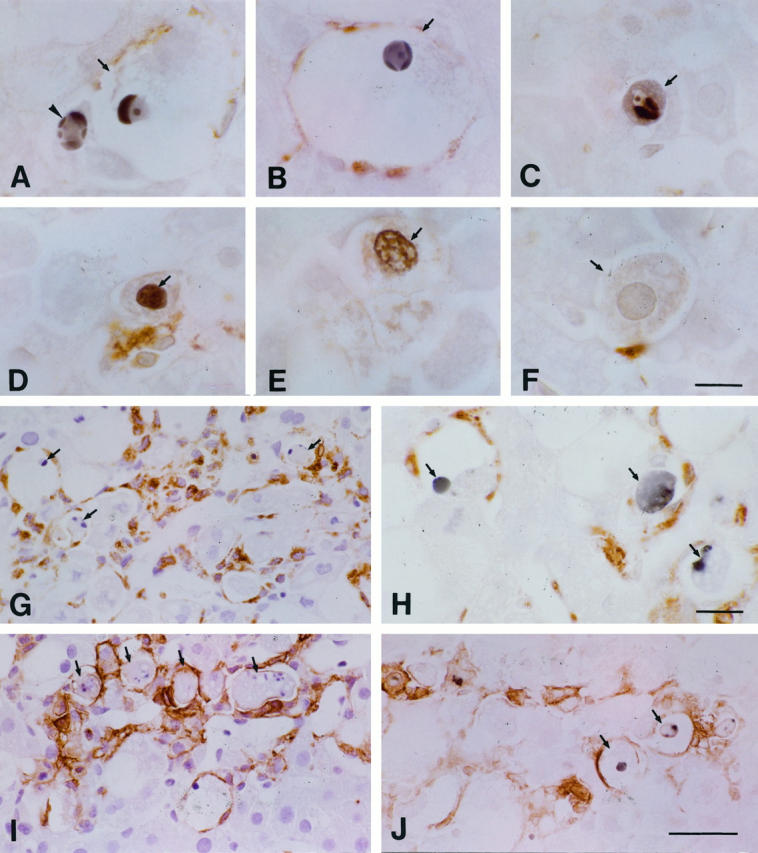
Immunostaining of the liver after the injection of CCl4. A and B: Hepatocytes with (arrows) and without (arrowhead) ballooned changes were identified both by TUNEL-positive staining and by their condensed chromatic masses or crescents. C: An apoptotic body (arrow) was seen in a hepatocyte. D and E: Hepatocytes (arrows) with TUNEL-positive staining but without highly condensed chromatic masses or crescents were present. F: A hepatocyte (arrow) appeared without TUNEL-positive staining (or its cytoplasm was only weakly and nonspecifically stained). G: Many apoptotic hepatocytes (arrows) were phagocytosed or were surrounded by ED1+ cells (brown). H: TUNEL+ cells (arrows) were surrounded by ED2+ cells (brown). I: Similarly to G, many of the apoptotic cells had incorporated with MHC class II Ag+ cells. J: TUNEL-positive cells (arrows) were surrounded by MHC class II Ag+ cells (brown). Bars: A to F, 10 μm; H, 15 μm; G, I, and J, 40 μm.