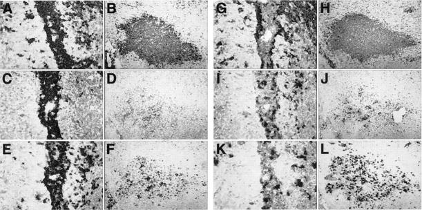
Figure 6.
Immunophenotypic characterization of inflammatory lesions in the cerebellum of a symptomatic GT-8 mouse. Immunohistochemistry was performed as described in Materials and Methods. Comparison of a perivascular (A, C, E, G, I, and K) with a parenchymal lesion (B, D, F, H, J, and L) immunostained for CD45 (A and B), B220 (C and D), CD4 (E and F), Mac-1 (G and H), CD8 (I and J), and neutrophils (K and L).

