
Figure 1.
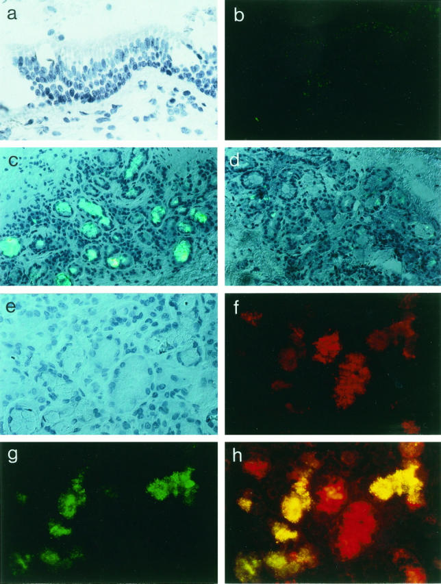
Expression and localization of chemokine IL-8 in human ΔF508 homozygous CF and non-CF bronchial tissues. Analysis of frozen tissue sections (5 μm thick) in CF bronchial surface epithelium (a and b) and submucosal gland structures from ΔF508 homozygous CF patients (c and e–h) and non-CF (control) patients is presented (d). Shown are Nomarski photomicrographs (a and e) and immunofluorescence micrographs for detection of IL-8 with FITC (green; c, d, and g) and lysozyme with Texas red (red; f and h). Note the absence of immunostaining for IL-8 at the level of ΔF508 homozygous CF bronchial surface epithelium (b) as in non-CF bronchial surface epithelium (not shown). In contrast, dense IL-8 staining is specifically observed in most of the submucosal gland cells from CF bronchial tissues (c) but is not identified in the submucosal glad cells from non-CF bronchial tissues (d). At higher magnification (×400), ΔF508 homozygous CF bronchial submucosal glands (e–h) are photographed using fluorescence filters for simultaneous red (lysozyme; f)/green (IL-8; g) visualization. The yellow color (h) indicates significant co-staining of IL-8 and lysozyme in ΔF508 homozygous CF bronchial submucosal gland serous-type cells. Results are representative of the findings in eight ΔF508 homozygous CF patients and four non-CF (control) subjects. All sections are counterstained with hematoxylin.