
Figure 2.
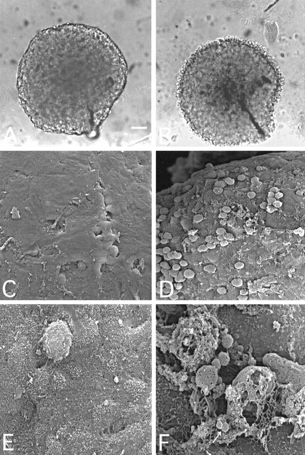
Light (A and B) and scanning electron (C to F) microscopic images of intact (A, C, and E) and C-attacked (B, D, and F) T47D breast adenocarcinoma cell spheroids. Spheroids were selected for the experiments after 10 to 14 days of culture and exposed to YTH53.1B and S2 and NHS at 37°C for 24 hours (B, D, and F) as in Figure 1B ▶ . The spheroid shown in A was photographed before the NHS exposure. In C and E, the spheroids were exposed to NHS, S0, and normal rat IgG for 24 hours. The surface of a spheroid exposed to C-activating (S2) and CD59-neutralizing (YTH53.1B) antibodies and NHS (B) was less coherent than that of the control (A). Surface cells became swollen and detached. Scanning electron microscopy also showed damaged, swollen, and detaching cells on the spheroid surface (D). At higher magnification, remnants of cells with destroyed, porous membranes could be seen with released vesicles (F, arrow). Bar, 100 μm (A). Magnification, ×500 (C and D) and ×3000 in E and F.