
Figure 3.
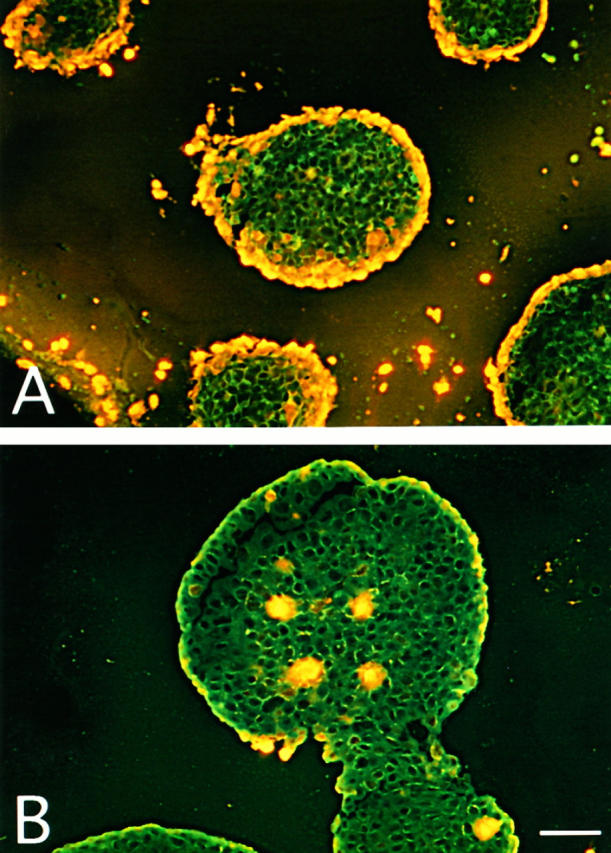
Visualization of C-mediated killing of PA-1 spheroids by the incorporation of PI into cellular DNA. A: PA-1 spheroids grown for 10 days were incubated with YTH53.1B (25 μg/ml) and S2 (50 μg/ml) antibodies and human serum for 4 hours at 37°C and with PI (50 μg/ml) for 1 hour. In the control (B), normal rat IgG (25 μg/ml) and S0 (50 μg/ml) were used instead of YTH53.1 and S2. After incubation, the spheroids were frozen in liquid nitrogen. Examination of cryostat sections (10 μm) by fluorescence microscopy shows PI staining of the nuclei of killed cells. In A, rims of dead cells can be seen on the surfaces of the spheroids. In the control (B), occasional necrotic cells in the center of a spheroid are stained. Bar, 200 μm (A).