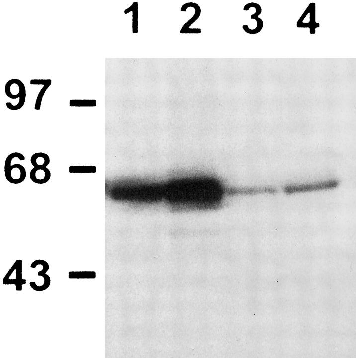
Figure 10.
Western blot analysis for p65 protein. Cytosolic extracts of type II alveolar cells freshly isolated from unexposed animals (lane 1) and from rats exposed for 7 days to 85% oxygen (lane 2). Nuclear extracts of type II cells isolated from unexposed control rats (lane 3), and from animals exposed to 85% oxygen (lane 4). Molecular weight markers in kd are shown to the left.

