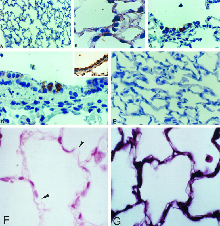
Figure 5.

Immunohistochemical localization of gelatinase B (MMP-9) in lung tissue sections. Immunoreactive gelatinase B was revealed by peroxidase reaction. Sections were lightly counterstained with hematoxylin. A: Normal lung (magnification, ×70). B to D: Lung of rat exposed 7 days to hyperoxia, at higher magnification (×350), showing alveolar (B and C) and bronchiolar (D) epithelial cells. D (inset) shows bronchiolar epithelial cells stained with cytokeratin. E: Control lung after 7 days of hyperoxia with primary antibody omitted (×70). F: Lung tissue from a rat exposed 7 days to hyperoxia, stained with anti-type IV collagen; arrows indicate areas of basement membrane disruption (×350). G: Normal lung tissue stained with anti-type IV collagen.
