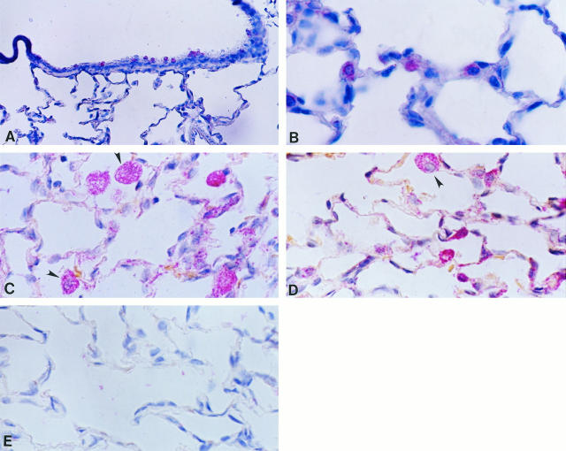
Figure 8.
In situ hybridization for TIMP-1 and TIMP-2 in controls and in 85% oxygen-exposed rats from 3 days. Tissue samples were processed for in situ hybridization with digoxigenin-labeled riboprobes and after that with anti-digoxigenin antibody coupled to alkaline phosphatase. The color reaction was performed with Fast Red chromogen, and tissues were slightly counterstained with hematoxylin. A: TIMP-1 expression in bronchiolar cells from a normal lung; original magnification, ×70. B: TIMP-2 expression in alveolar epithelial cells in a normal lung; ×210. C: TIMP-1 expression in lungs hyperoxic for 3 days; positive alveolar macrophages are indicated by arrowheads; ×210. D: TIMP-2 expression in rats exposed 3 days to hyperoxia, exhibiting label in macrophages (arrowhead) and alveolar epithelial and interstitial cells; ×210. E: Sense riboprobe; 140×.

