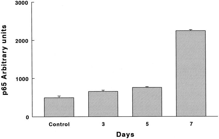
Figure 9.
NF-κB (p65) protein graph of immunoreactive protein. Lung tissue sections of normal and hyperoxia-exposed rats were treated with a polyclonal antibody directed against an epitope of the carboxy terminus of the Rel-A (p65) protein. Immunoreactive protein was revealed by peroxidase reaction. Quantification of Rel-A (p65) protein was performed with an image analyzer. P = 0.003, 0.0006, and 0.0005 at 3, 5, and 7 days, respectively.

