
Figure 3.
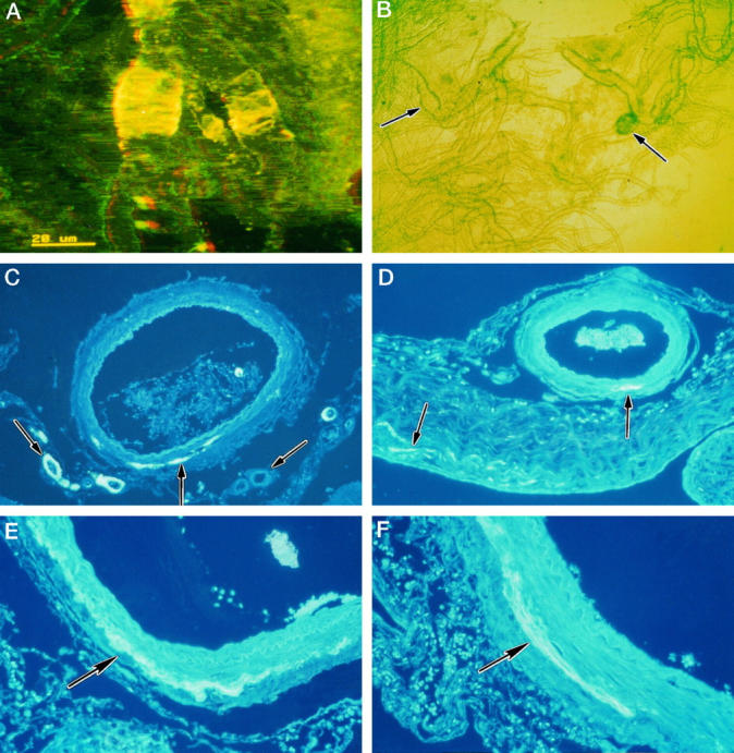
CAA of leptomeningeal arteries in AD. A: Isolated leptomeningeal artery showing heavy deposition of amyloid as transverse bands within its walls, associated with aneurysmal dilatation. Confocal microscopy: three-dimensional reconstruction, thioflavin S stain. B: Isolated leptomeningeal vessels viewed by phase-contrast microscopy showing a fusiform microaneurysm (left arrow) and a saccular microaneurysm (right arrow). C to F: Patterns of amyloid deposition in paraffin sections of leptomeningeal vessels and arachnoid stained with thioflavin S. C: Some smaller arteries show amyloid deposition throughout the thickness of their walls (left arrow), whereas other small vessels show complete absence of amyloid (right arrow). In the larger vessel, amyloid is deposited as a linear streak (middle arrow) in the adventitia and perivascular space. D: Leptomeningeal artery with a small streak of amyloid deposited at the junction of the media and adventitia (right arrow). The associated arachnoid mater also contains small streaks of amyloid (left arrow). E: Part of the wall of a leptomeningeal artery showing a linear circumferential deposit of amyloid at the junction of the adventitia and the media (arrow). F: Linear deposit of amyloid (arrow) in the wall of a leptomeningeal artery; the deposit is mostly in the adventitia near the perivascular space but also extends into the outer media.