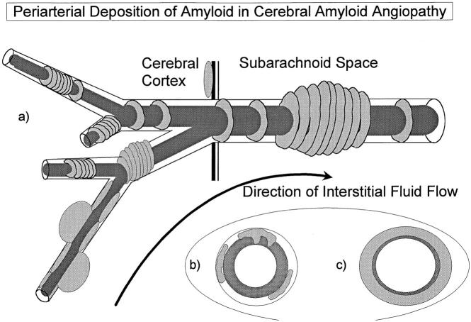
Figure 4.
Diagram summarizing the pattern of the distribution of amyloid in CAA associated with AD. a: Aβ accumulates as globules or linear deposits in the perivascular spaces of small intracortical blood vessels or as transverse bands in the walls of larger intracortical arteries and in smaller leptomeningeal arteries. The severity of amyloid angiopathy decreases with increasing size of the artery, suggesting that Aβ is precipitated to a greater extent in the initial portions of the pathways draining ISF from the brain. b: With increasing deposition, Aβ surrounds smooth muscle cells in the media. c: Eventually, smooth muscle cells are lost and aneurysms may form.

