
Figure 4.
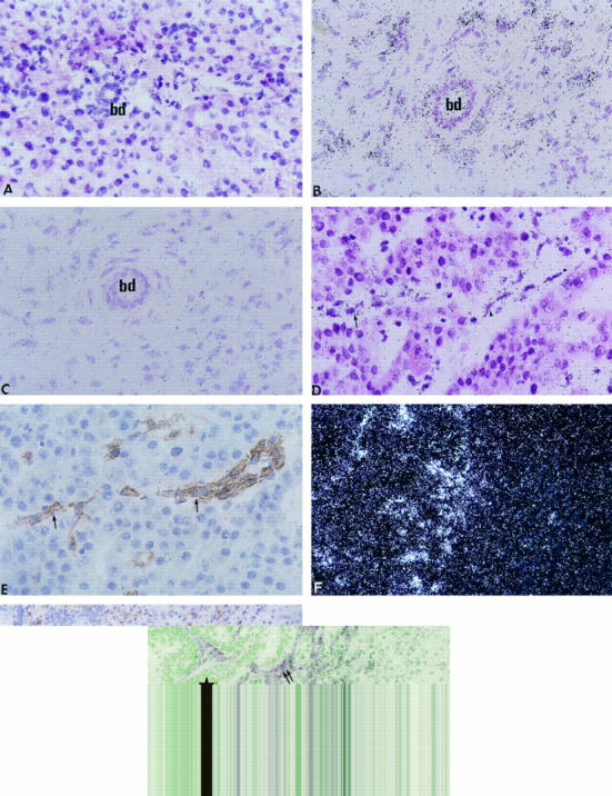
In situ hybridization of MT1-MMP mRNA in normal liver (A), cholestatic liver (B), HCC (D), and metastasis from a colonic adenocarcinoma (F). Controls include an immunohistochemical detection of von Willebrand factor in HCC (E, section contiguous to D) and α-smooth muscle actin in metastasis (G, section contiguous to F) and in situ hybridization with the sense probe (C, section contiguous to B). bd, bile duct; star, tumor; arrows, endothelial cells; double arrows, myofibroblasts. Bright-field (A to D) and dark-field (F) photomicrographs of autoradiographs stained with hematoxylin and eosin. Magnification: A to E, ×400; F and G, ×200.