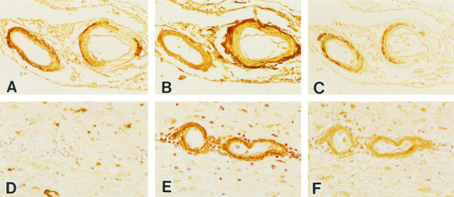
Figure 2.
Serial sections of leptomeningeal vessels (A–C) and parenchymal vessels (D–F) in the temporal lobe of an AD brain. These sections were stained with anti-Aβ (A and D), anti-AGE (B and E), and anti-ApoE (C and F) antibodies. Blood vessels with amyloid angiopathy are labeled by both anti-AGE (B) and anti-ApoE (C). Some blood vessels without amyloid deposits are labeled by both anti-AGE (E) and anti-ApoE (F). Magnification, ×125 (A–C) and ×200 (D–F).

