
Figure 6.
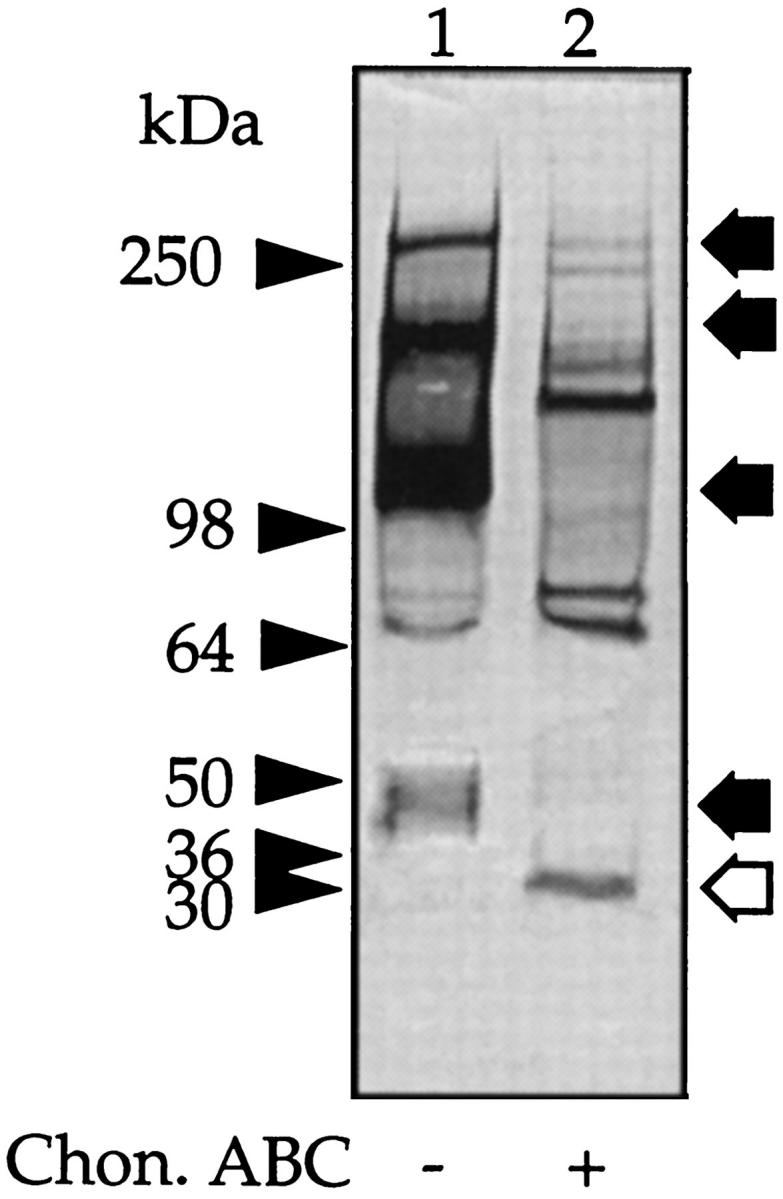
Western blot of peritoneal fluid. Peritoneal fluid (5 μl) was incubated with buffer alone (lane 1) or with chondroitin ABC lyase (lane 2), and a Western blot was generated with anti-human IαI antibody. The prestained molecular mass markers are indicated with arrowheads and the resolved PGs with arrows. The open arrows indicate the core protein released after incubation with chondroitin ABC lyase.