
Figure 2.
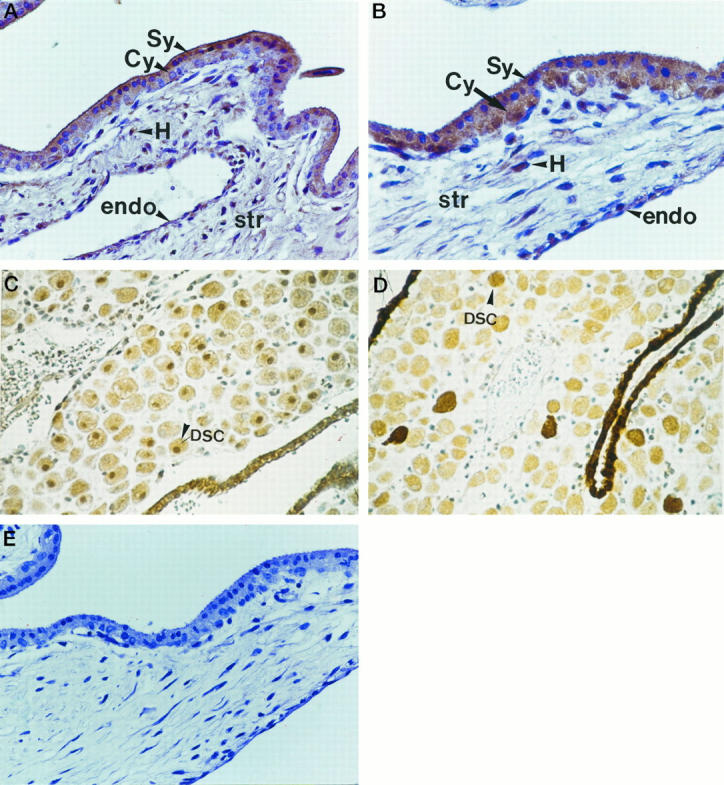
In the first trimester villus, the syncitiotrophoblast (Sy) and cytotrophoblast (Cy) layers of the villus were stained immunopositive to HGF (A) and c-met (B). The mesenchymal stromal core (str) was relatively loosely structured and stained positively for HGF along with the Hofbauer cells (H). The vascular endothelium (endo) stained weakly positive for both HGF (C) and c-met (D). Control sections (see text) were negative (E).