
Figure 3.
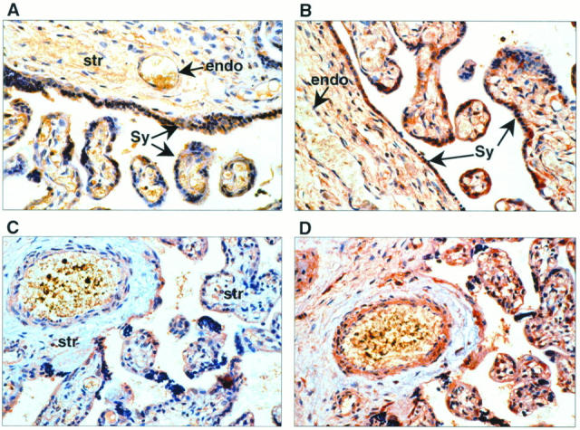
In placentae from uncomplicated third trimester pregnancies, positive cytoplasmic staining was identified using anti-HGF antibody (A), most intensely in the syncytium (Sy). There was also positive immunoreactivity to HGF in the endothelial cells (endo) lining the vasculature of the villi and the cells within the mesenchymal stromal core (str). B: c-met immunolocalization is primarily to the vascular endothelium (endo) within villi and to the syncytium (Sy) around the villi. In third trimester placentae complicated by IUGR, HGF immunoreactivity was localized in the same tissue types as in placentae of “uncomplicated” pregnancies (C). However, the HGF immunostaining intensity was less marked in the mesenchymal stroma. In serial sections immunostained for c-met, there was no discernible alteration in either localization or intensity (D).