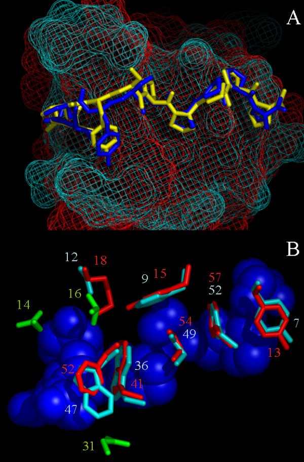
Figure 9.

The R21A Spc-SH3 and Abl-SH3 p41-binding interface. A. Stick representation of p41 in complex with R21A Spc-SH3 (p41 yellow, SH3 red mesh) and with Abl SH3 (p41 blue, SH3 cyan mesh). Structural differences are mainly around Tyr4 due to the larger side chain of its H-bond partner in SH3 (Lys18 in Spc, Ser12 in Abl). B. Residues of SH3 involved in binding (R21A Spc-SH3 red, Abl SH3 cyan, p41 blue). Residues 14, 16 and 31 in Abl-SH3 (colored green) show contacts (LIGPLOT analysis [24]) in Abl-SH3 that are not present in Spc-SH3.
