Figure 3.

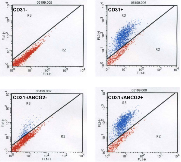
Flow analysis of prostate epithelial cells. Epithelial cells collected on a Percoll density gradient were stained with CD31 and ABCG2. Cells in R3 of each cytogram were sorted for further analysis.

Flow analysis of prostate epithelial cells. Epithelial cells collected on a Percoll density gradient were stained with CD31 and ABCG2. Cells in R3 of each cytogram were sorted for further analysis.