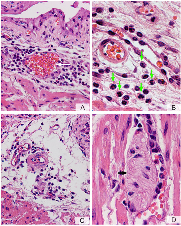
Figure 4.
A-D. PAR4-AP induces bladder inflammation. Anesthetized female C57BL6 mice were catheterized, the bladder was emptied, and a volume of 200 μl of a solution of PAR4-AP (10 μM) was instilled into the urinary bladder. Twenty four hours later, bladders were removed, processed for histology, and stained with H&E. A characteristic photomicrograph represents sub-urothelium inflammatory infiltrate around a blood vessel (A) and dilation of blood vessels (white arrow). At higher magnification (B), it was possible to visualize that the majority of inflammatory cells in response to PAR4-AP presented a characteristic "doughnut" shape indicative of mouse PMNs (green arrowhead). The submucosal edema is illustrated in C. Figure 4D illustrates inflammatory cells surrounding a structure resembling a nerve element (black arrow). Magnifications A = ×200, B = ×400, C = ×200, and D = ×400.

