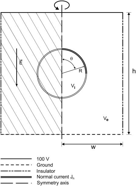
FIGURE 1.
Model of a cell in a uniform electric field. The model was mirrored through a symmetry axis for visualization (the mirrored portion is filled with a stripe pattern). The cell and the simulation region are not drawn to scale. Solutions for the potential inside (Vi) and outside (Ve) the cell determined using the Laplace equation subject to conditions on the boundaries listed above. The transmembrane potential difference, TMP, is the difference between Vi and Ve at the boundary.

