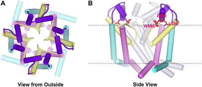
FIGURE 6.
Schematic representation of our model for hERG's pore-forming domain. Cylinders represent α-helices. Color code, S5 = cyan, S5-P linker = purple, P segment = yellow, S6 = pink. (A) View from the extracellular side through the pore. (B) Side view with two subunits colored by segment. The subunit at the back is colored light gray, and the subunit nearest the viewer has been removed. Highly conserved residues of the S5-P linker (W585, L586, and L589) are shown and colored red. Each S5-P linker is postulated to possess two helices (S5-P1 helix, H578-I583; and S5-P2 helix, W585-G594) that are connected by a hinge G584.

