
Figure 1.
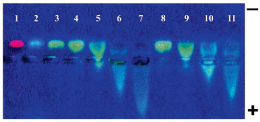
Effects of increasing SDS concentrations on gel electrophoresis of fluorescently labeled PVP-1300 and PVP-1300-coated SWNTs. Wells 1–3 PVP labeled with Alexa Fluor 594 (red fluorescence), Alexa Fluor 350 (blue fluorescence), and Alexa Fluor 488 (green fluorescence). Wells 4–7 SWNTS coated in green fluorescent PVP: well 4, 0.05% SWNT/PVP ratio (0.1% SDS); well 5, 0.1% SWNT/PVP ratio (0.2% SDS); well 6, 0.2% SWNT/ PVP ratio (0.4% SDS); well 7, 0.3% SWNT/PVP ratio (0.7% SDS). Wells 8–11 only fluorescent PVP and SDS no SWNTs: well 8, 0.1% SDS; well 9, 0.2% SDS; well 10, 0.4% SDS; well 11, 0.7% SDS. Amount of PVP in all wells was 35 μg/well. Note identical changes in the direction of movement of PVP-FL and PVP-FL SWNTs caused by SDS.