
Figure 5.
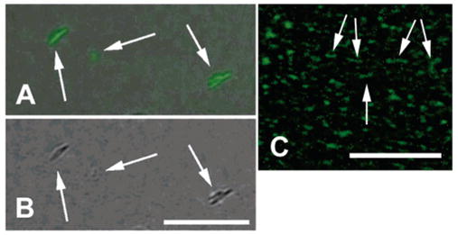
Fluorescing PVP-coated SWNTs deposited on a glass slide. (A) Combined light and fluorescent microscopy of nanotube bundles. Note a Y-junction on the right. (B) Light microscopy of the same bundles. Bar repesents 7 μm. (C) Individual fluorescent nanotubes are only seen using fluorescence detection. Note linear fluorescent nanotubes with the absence of junctions. Bar represents 4μm.