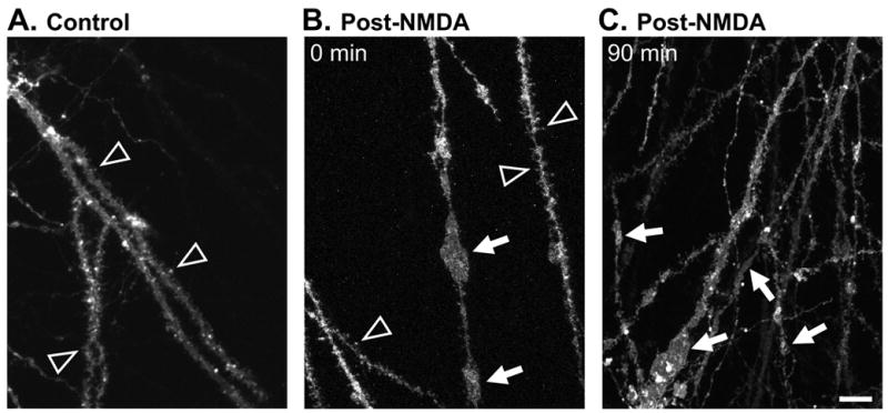
Figure 3. Persistence of irregular dendritic swelling visualized using DiO.

The lipophilic indicator DiO was loaded ballistically following slice fixation (see Methods) to label a small percentage of neurons. Images include regions in stratum radiatum centered ~100μm from stratum pyramidale. A: Representative control preparation showing dendritic spines (arrowheads) and no dendrite swelling. B: Preparation fixed immediately post-NMDA, showing some large irregular swellings (arrows) and some dendrite segments retaining fine dendritic spines (arrowheads). C: Preparation fixed 90 min after transient NMDA exposure, showing irregular swellings (arrows) in branches of dendrites with a range of diameters. Each image is representative of 5 slices analyzed per condition. Scale bar: 10μm.
