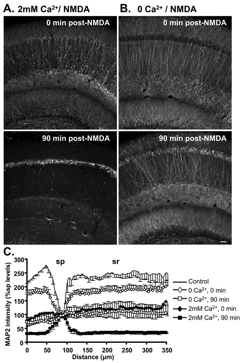
Figure 6. Ca2+ removal during NMDA exposure attenuated MAP2 redistribution.

MAP2 distribution following NMDA exposure in either 2mM Ca2+ (A) or Ca2+-free solution (B). Top panels are immediately following 10 min NMDA, and bottom panels are after 90 min washout (2mM Ca2+ present throughout washout in both cases). When Ca2+ was removed during NMDA exposure, redistribution of MAP2 immunofluorescence from dendritic to somatic compartments was substantially decreased immediately following exposure (top panels). At 90 min post-NMDA, some somatic MAP2 accumulation was still evident despite Ca2+ removal, but the loss of MAP2 from dendritic compartments was greatly decreased in slices exposed to NMDA in 0 Ca2+ (bottom panels). Slices in A&B were from the same animals, and processed in parallel. Scale bar (50μm) applies to all panels. C: Normalized MAP2 fluorescence intensity, plotted as a function of distance across slices (normalized against sp levels, mean±SEM from 5 experiments). Following stimulation in Ca2+-free solution (90 min, open squares), MAP2 immunofluorescence was relatively evenly distributed across the slice, in contrast to the predominant somatic accumulation observed in 2mM Ca2+ (90 min, filled squares).
