
Fig. 1.
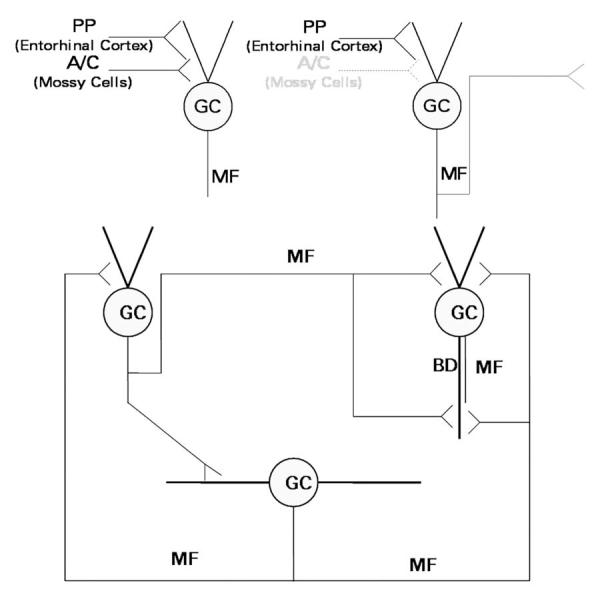
Schematic diagram of the excitatory innervation of dentate granule cells (GC). Upper left, In normal brain, granule cells receive excitatory innervation from the perforant path (PP), which originates in the entorhinal cortex, and the associational-commissural (A-C) fibers, which originate from mossy cells of the dentate hilus. Their axons, the mossy fibers (MF), innervate pyramidal cells of area CA3 and interneurons of area CA3 and the dentate gyrus, but only minimally innervate granule cells. Upper right, Seizures kill the hilar mossy cells, triggering the development of mossy fiber collaterals that grow into the synaptic territory abandoned by the degenerated associational-commissural fibers. Bottom, The granule cell network develops with time after brain-damaging seizures. Components of this network include normally-located granule cells of normal cellular morphology (upper left), normally-located granule cells with a basal dendrite (BD; upper right), and newly-generated hilar ectopic granule cells (bottom). These components are synaptically interconnected by mossy fibers. In animal models, these fibers express NPY de novo.