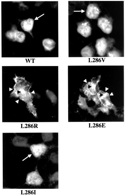
Figure 4.
Cellular distribution of Notch in cells expressing the indicated PS1 derivatives. After fixation in paraformaldehyde, cells were permeabilized and stained with antibody 9E10 to the cytoplasmic myc tail of NotchΔE. As observed before (22), cells expressing wt PS1 show an accumulation of Notch epitopes (most likely NICD) within the nucleus (arrows). In contrast, cells expressing PS1 L286E or PS1 L286R accumulate Notch epitopes at the plasma membrane (white arrowheads) and within the Golgi (46) (black arrowheads).

