
Fig. 3.
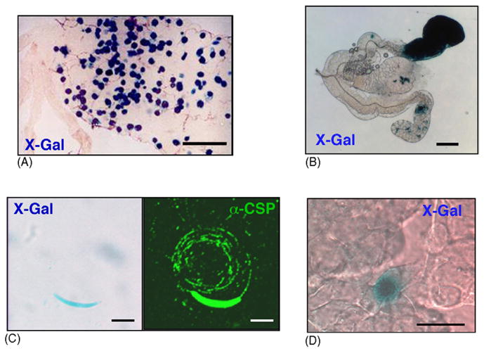
Detection of transgenic sporogonic and pre-erythrocytic Plasmodium stages expressing β-galactosidase. (A) Midgut oocysts of infected Anopheles mosquitoes at day 13 post feeding. All oocysts can be stained with X-Gal after fixation and permeabilization. Scale bar is 200 μm. (B) X-Gal staining of an infected salivary gland after permeabilization. Scale bar is 100 μm. (C) Transgenic sporozoites express β-galactosidase (left panel) and perform gliding locomotion indistinguishable from WT sporozoites (right panel). Scale bar is 5 μm. (D) Infected heptocytes can be stained with X-Gal after fixation and permeabilization. Shown is a 48 h liver stage. Scale bar is 20 μm.