
Fig. 6.
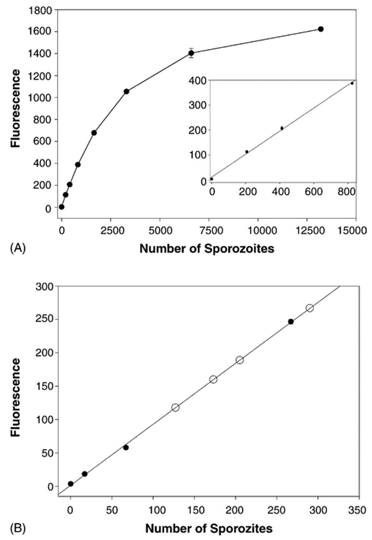
Quantification of PbBlue sporozoites injected into a live mouse by infected mosquitoes. (A) The fluorescent substrate 4-methylumbelliferyl-β-D-galactoside was used to detect PbBlue sporozoites dissected from salivary glands of infected mosquitoes. The inset shows the lower end of the curve in more detail. Shown is the mean with standard deviations of duplicates. (B) Different numbers of PbBlue sporozoites were added to homogenized mouse skin and a standard curve was generated (black circles). Infected mosquitoes (10–15) were allowed to probe on a mouse’s ear for 5 min, the ear was removed and processed as outlined in Section 2. Shown are the results from four ears (open circles) which are plotted on the standard curve generated above. The number shown is 1/15th of the total number deposited by the mosquitoes since only this fraction of the lysate was analyzed.