
Figure 6. Time kinetics of ERK1/2 activation in the lateral amygdala.
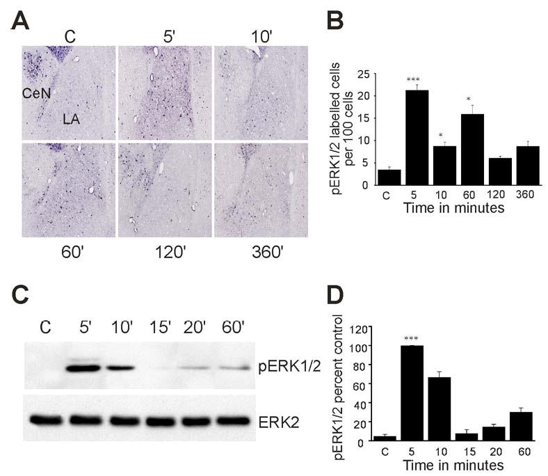
Rats were presented with two pairs of tone and shock and processed for immunohistochemistry or immunoblotting. (A) Representative immunohistochemical staining of pERK1/2 in the amygdala at 0, 5, 10, 60, 120 and 360 min after fear conditioning. (B) Bar diagram showing pERK1/2 immunoreactive cells per 100 cells in serial sections through the LA (mean ± S.E., n = 5 sections per time point). *p < 0.05, **p < 0.01, ***p < 0.001 relative to control. (C) pERK1/2 immunoblot of tissue punches taken from the LA at 5, 10, 15, 20 and 60 min after fear conditioning training (upper panel). The blots were reprobed with total ERK2 antibody (lower panel). (D) Quantification of pERK1/2 levels from 3 separate experiments (***p < 0.001).