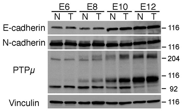
Figure 1.

Immunoblot of E-cadherin, N-cadherin and PTPμ in the developing chick retina. Lysates from nasal or temporal retina were prepared from E6, E8, E10 and E12 chicks, separated by SDS-PAGE, transferred to nitrocellulose membrane, and probed with an antibody to E-cadherin, N-cadherin or PTPμ (SK18). E-cadherin protein migrates at ~120 kDa, while N-cadherin migrates at ~130 kDa. Full length PTPμ is ~200 kDa whereas the proteolyticaly processed form of PTPμ containing the cytoplasmic domain migrates at ~100 kDa (Brady-Kalnay and Tonks, 1994). A 95 kDa immunoreactive band is also present. Each immunoblot was stripped and reprobed with antibodies against vinculin to verify equal protein load.
