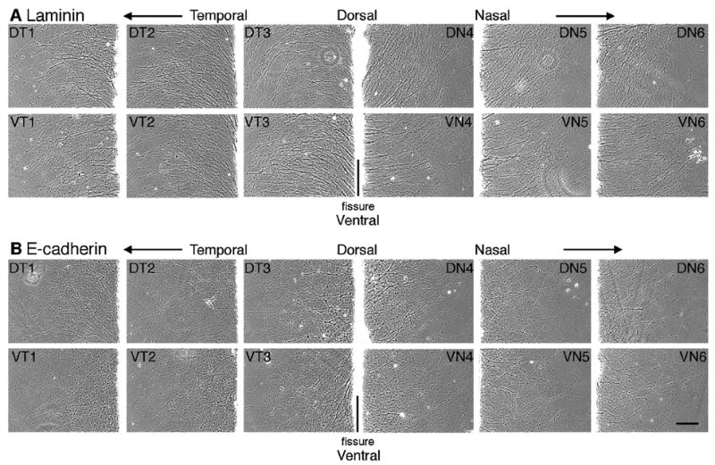
Figure 5.

Neurite outgrowth on E-cadherin and laminin is independent of RGC cell body origin. Explants from E8 chick retina were cut parallel to the optic fissure and explants from retina were cultured on E-cadherin (B) or laminin (A) substrates. Images were acquired after 20 hours in culture from a location corresponding to the outer third of each explant. Each number indicates the explant number (e.g. 1 and 6 are most peripheral). Dorsal (D), ventral (V), nasal (N), temporal (T). Scale bar, 200 μm.
