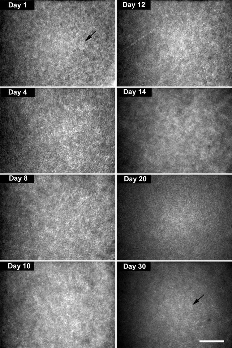
FIGURE 2.

Light-scattering detected by in vivo CM suggested the presence of densely packed ovoid or elliptical cell bodies (day 1, arrow). At day 14, smoother contours of individual cells could be detected (day 14). At day 30, faint outlines of more sparsely packed cells were detected (day 30, arrow). Bar, 100 μm.
