FIGURE 5.

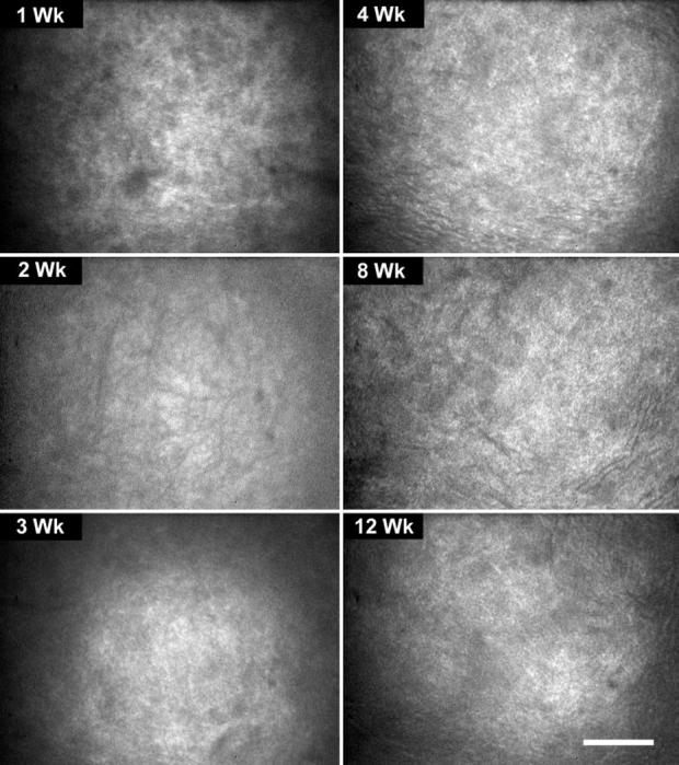
In vivo confocal microscopic images from the lumican-deficient mouse from 1 to 12 weeks of age showing a diffuse rather than granular light-scattering pattern, compared with the wild-type mouse. Bar, 100 μm.

In vivo confocal microscopic images from the lumican-deficient mouse from 1 to 12 weeks of age showing a diffuse rather than granular light-scattering pattern, compared with the wild-type mouse. Bar, 100 μm.