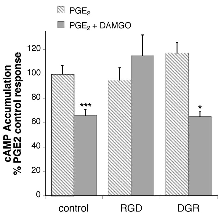
Figure 6.

RGD prevents inhibition of cAMP accumulation by DAMGO in BK primed TG cells. TG primary cultures (5–6 DIC) were incubated with or without soluble RGD peptides or reverse (inactive) sequence DGR peptide for 30 min (37°C) followed by addition of bradykinin (10 μM) and further incubation for 15 min (37°C). After pretreatment, cells were incubated with the phosphodiesterase inhibitor, rolipram (10 μM) with or without the MOR agonist, DAMGO (1 μM) and incubated for 15 min (37 °C) followed by addition of PGE2 (1 μM) and further incubation for 15 min (37°C). Cellular cAMP levels were determined by RIA. Data shown are expressed as the percentage of PGE2 stimulation in the presence of bradykinin and are the mean ± SEM of 3–5 experiments The presence of peptides did not alter either basal or PGE2 stimulated cAMP levels. Statistical analysis by one-way ANOVA (F(5,46) = 9.287) followed by Tukey’s post-test. * p < 0.01 comparison between PGE2 and PGE2+DAMGO for each pretreatment condition
