
Figure 1.
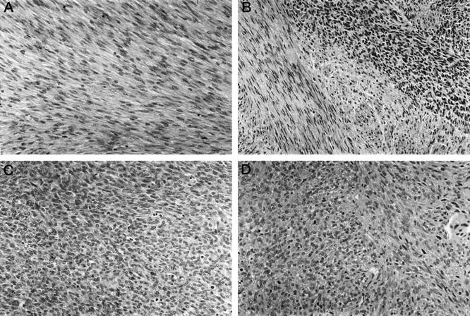
H&E-stained sections of classic CMN (A), mixed CMN (B), and cellular CMN (C) and CFS (D) are shown. The classic CMN consists of a moderately cellular proliferation of interlacing bundles of spindle cells whereas the cellular CMN exhibits a more densely cellular histology with increased mitotic activity. The mixed CMN contains a mixture of the two patterns. The CFS is very similar in appearance to the cellular CMN.