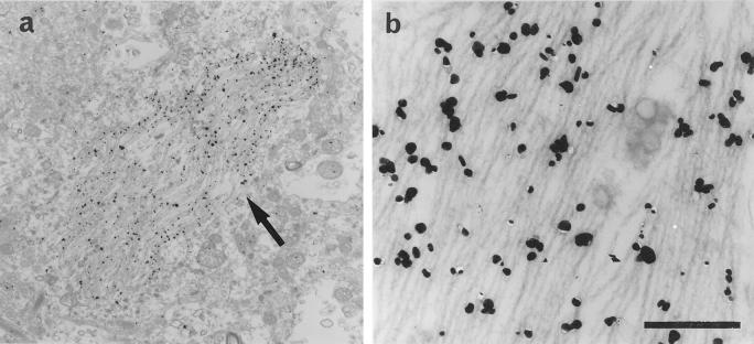
Figure 4.
Immunoelectron micrographs for Nedd5 in the entorhinal cortex of an AD brain. a: An electron micrograph showing that gold particles for Nedd5 (arrow) specifically labeled NFTs in the neuronal cytoplasm. Scale: 4 μm. b: A higher magnification of the NFTs. The gold particles are concentrated along PHFs in the cytoplasm. Scale: 0.4 μm.

